Minirhizotron Systems for Root Disease Detection
Dr. Vijayalaxmi Kinhal
January 8, 2024 at 6:46 pm | Updated April 15, 2026 at 2:36 am | 5 min read
- Root diseases are a significant issue in reducing crop yields globally.
- The damage and symptoms caused by root diseases change morphology and growth dynamics that images and scans can detect.
- Minirhizotron systems can detect a wide range of root changes reflecting various levels of root disease severity, providing a novel detection method.
Root diseases are a significant source of biotic stress that can harm roots. Without a functional root system, plants can grow stunted or result in total plant loss. However, detecting infected roots is not easily accessible due to the underground placement of these plant organs. Minirhizotron systems, including root imagers used through fixed transparent tubes, can provide a window into underground dynamics and root-pathogen interactions. Learn more about this new development that is helping research and precision agriculture.
Current Disease Detection Methods
Plant roots make up 30–95 percent of plant biomass. Roots functions are crucial for plant establishment, growth, and productivity. They anchor the plants, search and transport water and nutrients, and store biomass.
Root pathogens are a scourge of all crop species, as they cause a wide range of damage to roots, from mild to complete loss of roots. Common pathogen species affecting roots globally can be bacteria, fungi, viruses, etc., and essential groups are:
Subscribe to receive our monthly round-up of articles.
- Pythium that causes root rot in many crops.
- Verticillium is a fungus that attacks the vascular system, leading to wilt and plant death.
- Fusarium solani species complex (FSSC) causes Fusarium root rot in critical staple crops like potatoes, common beans, soybeans, melon, peas, and zucchini.
- Parasitism by broomrape (Orobanche cumana), a chlorophyll-less obligatory root parasite. They grow on roots for all their needs and can severely reduce yield in many crops, like legumes, sunflowers, tobacco, tomatoes, carrots, etc. In some cases, Orobanche is pathogenic in its effects. It causes diseases by interfering with hormonal balance and crop photosynthetic machinery.
Visual detection of symptoms helps growers to spot and treat infections in aboveground parts like leaves, branches, and stems. However, the underground nature of roots makes early disease detection more difficult. Therefore, loss to perennial orchard crops can be devastating, as disease can spread in infected roots, and the whole tree can be affected or die.
Current root evaluation methods for pathogens have several problems.
- The methods are destructive. The roots are removed from the soil and examined visually. Washing of roots can remove fine roots so that only 60 percent are examined during manual inspection. Or the soil is scrapped back to access tree roots and check root surface conditions.
- Methods provide a one-time snapshot of root conditions. Single observations do not shed any light on changes to root morphology and growth dynamics due to disease infections.
- Moreover, the system to classify severity is also not accurate. Disease scoring systems use semi‐quantitative scales, only visual symptoms, and miss several damages and variations caused to the roots’ morphology and functioning.
Taking image scans in combination with minirhizotrons can solve several issues with current root disease detection.
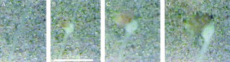
Figure 1: Changes in individual Orobanche cumana parasitism of a root over time: (A) 14 d after planting (DAP); (B) 21 DAP; (C) 28 DAP; (D) 35 DAP.
Root Traits for Disease Detection
Pathogen activity changes the root morphology, which image scanners used in a minirhizotron system can measure. For example, symptoms like necrosis or stunting change morphological root traits and can help their detection. The root traits helpful in this context can be any of the following:
- Discoloration of roots, for example, due to wilts, rot, galls, and cankers.
- Total root length estimations record root growth and dynamics as affected by diseases.
- Dead roots or root portions due to necrosis
- Volumetric growth of roots can decrease due to disease infection. For example, taproot growth is affected fourteen days after the start of a Cercospora
- Lateral root development changes can occur, for example, due to cyst nematodes in sugar beet.
- Deformation of tubers due to cyst nematode infection for four weeks is detectable after infection.
- Root infestation by broomrape parasites can be spotted by root scans, for example, in sunflowers, see Figure 1.
- Fine-root dynamics changes in response to plant protection measures.
The root traits listed above show the importance of continuously monitoring roots to detect disease development and incidence. This is why minirhizotron systems are valuable in disease detection. Moreover, the symptoms detected by the root traits reflect different disease stages and levels of severity, see Figure 2.
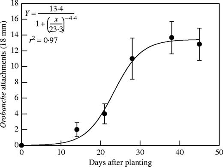
Figure 2: “Time course of Orobanche cumana parasitism in sunflower, quantified by means of minirhizotron. Measurements were taken at 20 cm depth. Bars represent the standard error of ten measurements” Eizenberg et al. 2005. (Image credits: https://doi.org/10.1093/aob/mci252).
Minirhizotron Systems
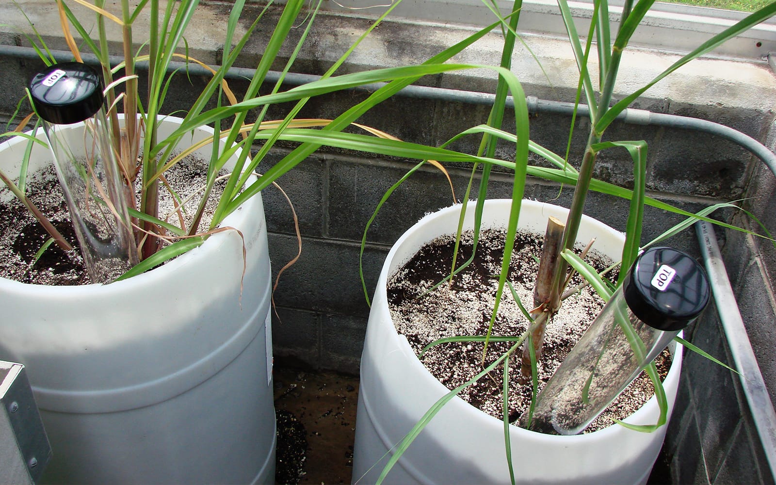
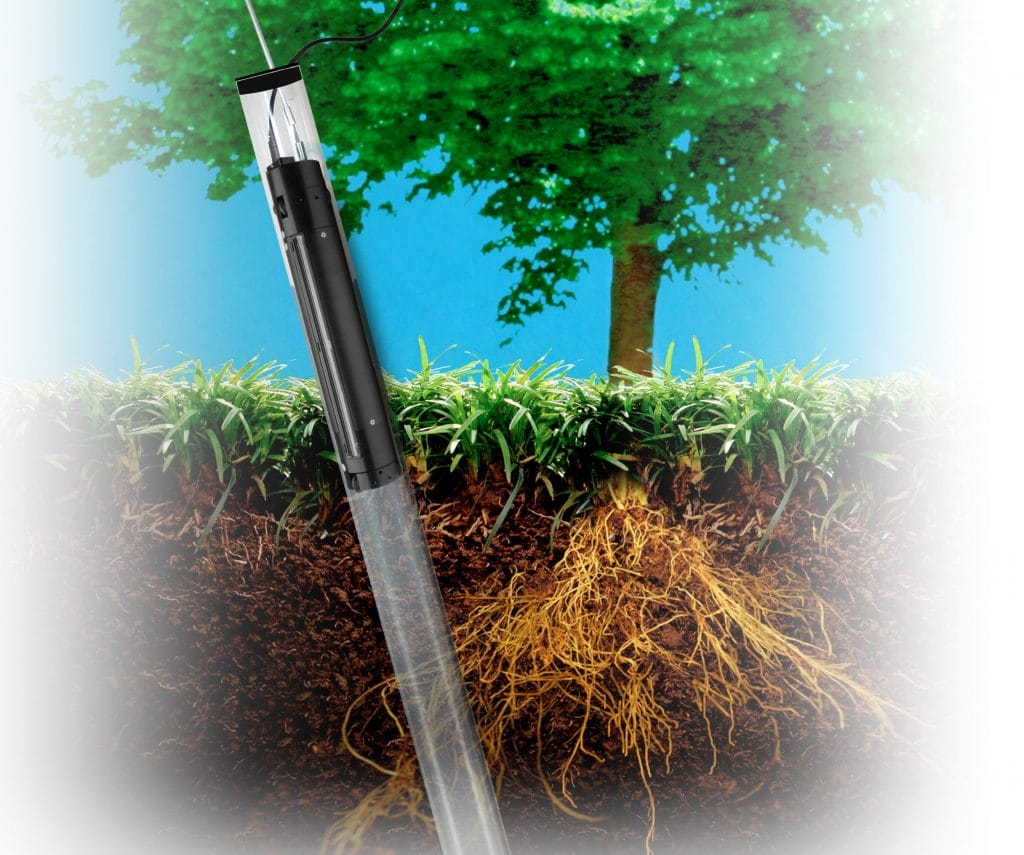
Minirhizotrons are transparent long tubes inserted into the soil at an angle, enabling the recording of a root system without disturbing the roots in any way after the initial installation. Since the tubes are left in the soil, long-duration experiments with repeated scans of roots at the soil-tube interface by a camera inserted can take high-resolution images of the roots growing around the tube. It is suitable for evaluating root morphology, growth, senescence, damage, and elongation. Data can be recorded often as the method is non-destructive, providing an accurate picture of changes occurring within days to months as required. The high-resolution images allow for the estimation of even fine-root growth and turnover.
In some cases, the images captured by the camera or scanner were still analyzed manually. This is a time-consuming process; results depend on image quality, roots, and abundance, and are not objective. For example, root rot was scored from 1 to 3, and significant quantities of missing roots were scored from 7 to 9. Manual evaluation of images is more suited for larger root systems than fine roots and can suffer from human error.
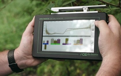
Therefore, nowadays, software-aided computer-based image analysis is becoming more common. The software uses deep learning AI models, such as neural networks, for automated and more accurate measurements of root traits like length, width, area, volume, count, etc.
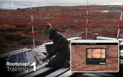
CID Bio-Science Imager Scanners
CID Bio-Science produces two root image scanners, the CI-600 In-Situ Root Imager and the CI-602 Narrow Gauge Root Imager, which have been used by scientists in experiments for non-destructive detection of fungal infection, nematode cysts, and parasites. The accompanying software RootSnap! makes minirhizotron systems image analysis easy. Both images are suitable for field experiments and precision agriculture. Since root tubes are inexpensive components, several can be installed in a field and used with root imagers to detect soil-borne diseases in large areas better.
Sources
Eizenberg, H., Shtienberg, D., Silberbush, M., & Ephrath, J. E. (2005). A New Method for in-situ Monitoring of the Underground Development of Orobanche cumana in Sunflower (Helianthus annuus) with a Minirhizotron. Annals of Botany, 96 (6), 1137–1140, https://doi.org/10.1093/aob/mci252
Fernández-Aparicio, M., Reboud, X., & Gibot-Leclerc, S. (2016). Broomrape weeds underground mechanisms of parasitism and associated strategies for their control: A Review. Frontiers in Plant Science, 7. https://doi.org/10.3389/fpls.2016.00135
Głuszek, S., Sas Paszt, L., Sumorok, B., Derkowska, E., & Kozera, R. (2013). Application of the MINIRHIZOTRON technique to studying the roots of Fruit Plants. Advances in Science and Technology – Research Journal, 7(18), 45–53. https://doi.org/10.5604/20804075.1049605
Peters, B., Blume-Werry, G., Gillert, A., et al. (2023) As good as human experts in detecting plant roots in minirhizotron images but efficient and reproducible: the convolutional neural network “RootDetector.” Sci Rep 13, 1399. https://doi.org/10.1038/s41598-023-28400-x
Pierz, L. D., Heslinga, D. R., Buell, C. R., & Haus, M. J. (2023). An image-based technique for automated root disease severity assessment using PlantCV. Applications in plant sciences, 11(1), e11507. https://doi.org/10.1002/aps3.11507
Tang, W., Wu, N., Xiao, Q., Chen, S., Gao, P., He, Y., & Feng, L. (2023). Early detection of cotton verticillium wilt based on root magnetic resonance images. Frontiers in Plant Science, 14. https://doi.org/10.3389/fpls.2023.1135718
Related Products
Most Popular Articles
- Transpiration in Plants: Its Importance and Applications
- Leaf Area – How & Why Measuring Leaf Area…
- How to Analyze Photosynthesis in Plants: Methods and Tools
- Plant Respiration: Its Importance and Applications
- The Forest Canopy: Structure, Roles & Measurement
- Stomatal Conductance: Functions, Measurement, and…
- Forest & Plant Canopy Analysis – Tools…
- Root Respiration: Importance and Applications
- 50 Best Universities for Plant Science
- The Importance of Leaf Area Index (LAI) in…