Watching Wetland Soils Change in Real Time: Auburn University’s Innovative Use of the CI-600 Rhizosphere Camera
Hunter Weber
October 22, 2025 at 10:20 pm | Updated October 22, 2025 at 10:20 pm | 3 min read
Understanding how soils transition between oxidized and reduced states is essential for accurately classifying and protecting wetlands. Traditional techniques such as the Indicator of Reduction in Soils (IRIS) method provide valuable data. Still, they rely on periodic sampling and the destruction of multiple test tubes, limiting both temporal resolution and sustainability.
Researchers at Auburn University have demonstrated a new way forward. By adapting the CI-600 Rhizosphere Camera, generally used for root imaging, they created a fully automated system that monitors soil redox dynamics in real time. Their proof-of-concept experiment offers a non-destructive, continuous alternative to the standard IRIS approach and reveals how reduction processes unfold hour by hour inside wetland soils.
From Manual Sampling to Real-Time Redox
Wetland soils are naturally low in oxygen. Measuring that lack of oxygen, the soil’s reduction-oxidation or “redox” status, is key to verifying whether a site qualifies as a wetland. For years, researchers have used the Indicator of Reduction in Soils (IRIS) test to do this.
Subscribe to the CID Bio-Science Weekly article series.
By submitting this form, you are consenting to receive marketing emails from: . You can revoke your consent to receive emails at any time by using the SafeUnsubscribe® link, found at the bottom of every email. Emails are serviced by Constant Contact
The IRIS method involves coating plastic tubes with iron or manganese oxides, inserting them into soil, and retrieving one tube each week to see how much coating has been removed by microbial activity. The more oxide removed, the more reducing (and wetter) the soil is.
The downside? Each test destroys multiple tubes and only gives a few snapshots in time.
Auburn’s team, led by Olivia LeFevre and Dr. Thorsten Knappenberger, set out to change that. They wondered if they could automate the process, capturing continuous data from a single, reusable tube. The answer came from an unlikely place: a root imaging camera.
Turning a Root Camera into a Soil Chemistry Lab
Originally designed to study plant roots, the CI-600 Rhizosphere Camera captures detailed 360° images inside transparent tubes installed in the soil. LeFevre and Knappenberger realized that with a few modifications, the camera could record not roots, but chemical reactions.
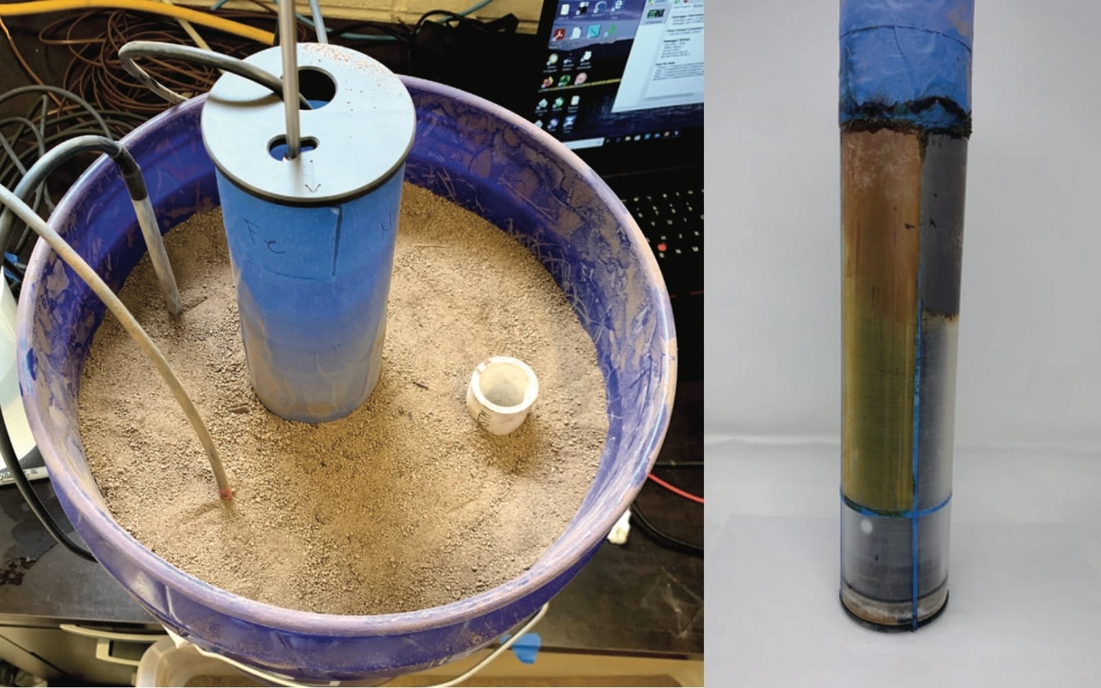
They coated a clear rhizosphere tube with alternating stripes of iron and manganese oxides and inserted it into a soil column packed with wetland soil and peat. Using a custom Python automation script, they programmed the CI-600 to take photos every hour for 28 days, while sensors continuously logged redox potential, temperature, and pH.
What the Camera Saw
Within the first 8 hours, manganese oxide began to reduce, showing visible changes on the coating. Iron oxide followed after about 4 days. Over time, the team tracked how the reduction progressed across different sections of the tube:
-
30% manganese reduction was reached in just 4.9 days
-
Iron reduction took roughly 9 to 13 days, depending on the depth
-
The maximum manganese reduction rate hit 20% per day
By the end of the experiment, some coatings were entirely gone, proof that the camera was capturing the microbial respiration and chemical shifts that define wetland soils.
Why It Matters
This new approach eliminates the need to pull and discard multiple IRIS tubes. Instead, one clear, reusable tube provides continuous, non-destructive data with precision that manual sampling can’t match.
For environmental scientists, engineers, and wetland consultants, the implications are significant:
-
Sustainability: Far less plastic waste compared to traditional IRIS methods.
-
Efficiency: Hourly data collection without field visits.
-
Accuracy: Consistent measurements from the same soil volume, reducing spatial variability.
-
Scalability: Potential for field use with portable or lower-cost imaging setups.
The study demonstrates how imaging and automation can streamline wetland verification and environmental monitoring—two processes that are often time-consuming and resource-heavy.
Looking Ahead
As Dr. Knappenberger noted, this proof-of-concept could eventually integrate with machine learning algorithms to automatically classify reduced areas, calibrate for different soil types, and even generate predictive models of wetland development.
For CID Bio-Science, it’s a compelling example of how the CI-600l, a tool built initially for studying roots, can expand into new areas of environmental research. When precision imaging meets soil chemistry, even the slow work of microbes becomes visible, measurable, and, for the first time, continuous.
Reference
LeFevre, O.V., Knappenberger, T., Shaw, J.N., & Olshansky, Y. (2021). Camera illustration of Indicator of Reduction in Soils (IRIS) reduction dynamics. Agricultural & Environmental Letters, 6:e20051.
https://doi.org/10.1002/ael2.20051
Related Products
Most Popular Articles
- Transpiration in Plants: Its Importance and Applications
- Leaf Area – How & Why Measuring Leaf Area…
- How to Analyze Photosynthesis in Plants: Methods and Tools
- Plant Respiration: Its Importance and Applications
- The Forest Canopy: Structure, Roles & Measurement
- Stomatal Conductance: Functions, Measurement, and…
- Forest & Plant Canopy Analysis – Tools…
- Root Respiration: Importance and Applications
- The Importance of Leaf Area Index (LAI) in…
- 50 Best Universities for Plant Science