CI‑600 In‑Situ Root Imager vs Traditional Minirhizotron: Time, Labour & Insights
Scott Trimble
November 18, 2025 at 5:56 pm | Updated November 18, 2025 at 5:56 pm | 4 min read
The CI-600 In-Situ Root Imager is a modern solution for non-destructive root observation and data collection. Compared to traditional minirhizotron systems, it delivers faster image capture, greater consistency, and richer insight into below-ground dynamics while cutting the time and labour typically associated with root research.
The Challenge of Root Observation
Roots drive plant performance, yet remain the hardest part of a plant to study. Conventional minirhizotron systems, often custom-built or manually assembled, require bulky cameras, limited field mobility, and a tedious workflow involving photo stitching, calibration, and data correction.
The CI-600 In-Situ Root Imager by CID Bio-Science solves these long-standing issues by automating image collection in a compact, field-ready design that prioritizes accuracy, repeatability, and researcher efficiency.
Subscribe to the CID Bio-Science Weekly article series.
By submitting this form, you are consenting to receive marketing emails from: . You can revoke your consent to receive emails at any time by using the SafeUnsubscribe® link, found at the bottom of every email. Emails are serviced by Constant Contact
What Makes the CI-600 In-Situ Root Imager Stand Out
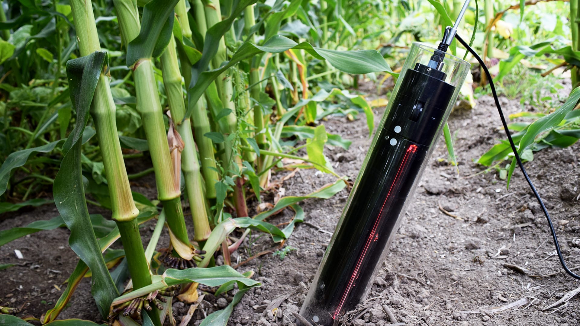
The CI-600 captures high-resolution, panoramic images of roots growing against transparent tubes installed in the soil. It replaces the mechanical complexity of older minirhizotron setups with a single, motorized scanner that rotates inside the tube, capturing a complete 360° view in one pass.
Key Specifications and Capabilities:
-
Scan Window Length: 9 1/8″
-
Full 360° Image Capture with no need for manual rotation
-
High-resolution, non-destructive imaging of living roots
-
Compatible with CID RootSnap! software for automatic image analysis
-
Lightweight, portable design ideal for field studies
Together, these features allow researchers to visualize and quantify root growth patterns, branching, and turnover without disturbing the plant or soil structure.
Time Efficiency: From Hours to Minutes
Traditional minirhizotron systems often require several steps: manual tube alignment, camera adjustment, focus calibration, and multiple partial scans stitched together in post-processing. The CI-600 streamlines this entire process.
-
Single-Scan Operation: The imager rotates automatically, capturing the full circumference of the tube in seconds.
-
Instant Image Storage: Data is saved directly to internal memory and can be analyzed on-site.
-
No Manual Stitching: Images are seamlessly assembled into panoramic root maps by the RootSnap! software.
In practice, this cuts total imaging time per sample from 30–60 minutes to less than five minutes, allowing far larger data sets to be collected across experimental plots.
Reducing Labour in Root Studies
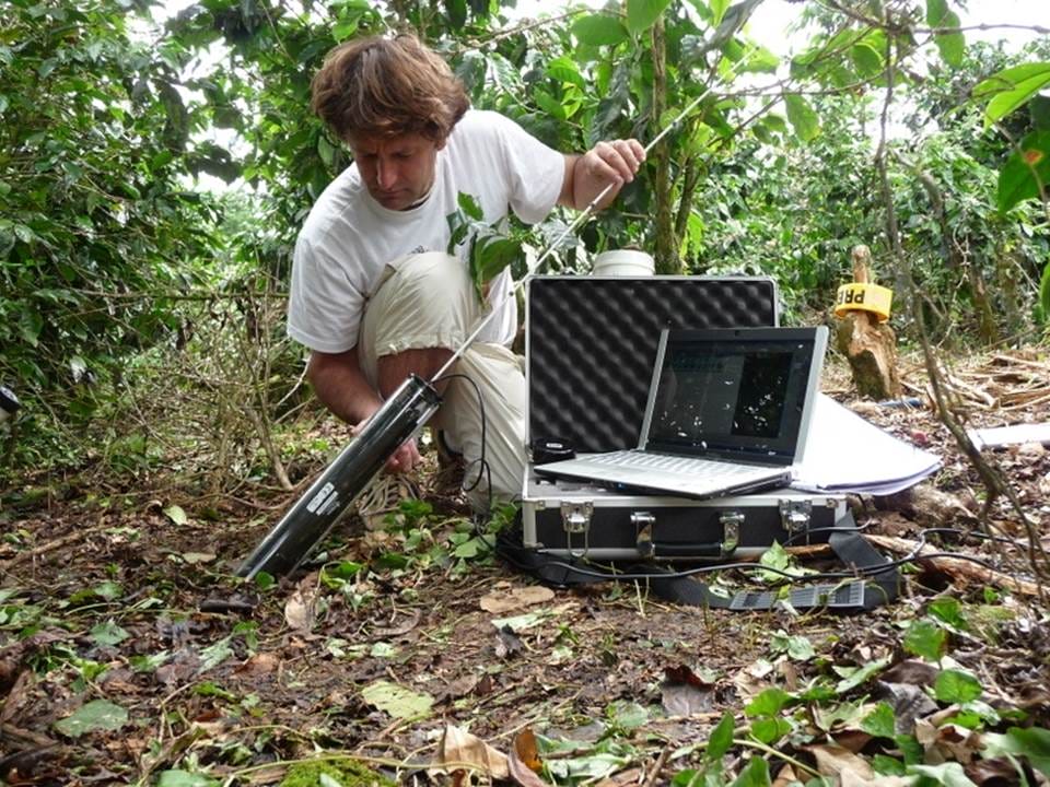
Root phenotyping has historically been labour-intensive, often requiring multiple technicians per site to handle camera equipment, laptops, and calibration tools.
The CI-600 In-Situ Root Imager reduces that burden with:
-
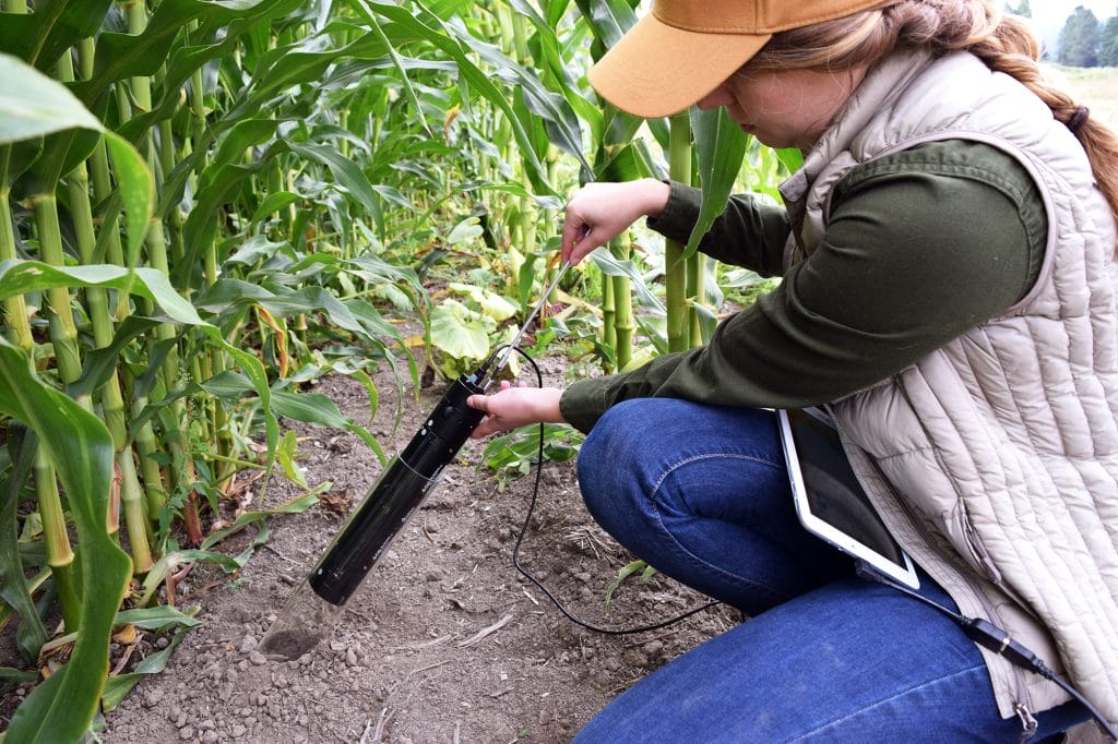
Single-operator usability, allowing one person to image multiple tubes efficiently
-
No above-ground setup or alignment, as the imager’s motorized scan ensures consistent positioning
-
Automated analysis tools, with integration through CID’s RootSnap! software that measures root length, diameter, area, and branching patterns automatically
Fewer manual tasks mean researchers can focus on experimental design and data interpretation rather than repetitive imaging work.
Data Quality and Research Insights
While traditional minirhizotrons can produce decent imagery, they often struggle with focus uniformity and depth accuracy. The CI-600 solves these issues by using precision optics and consistent illumination along the tube’s length.
Advantages in Image Quality:
-
Uniform lighting and resolution across the entire 360° field
-
Non-destructive imaging that preserves the natural soil environment
-
Repeatable positioning for longitudinal studies over months or seasons
Insights Enabled by the CI-600:
-
Root growth rates in response to drought, nutrient treatments, or soil compaction
-
Root turnover and mortality patterns in perennial crops and trees
-
Fine root development across soil horizons
-
Species-specific root architecture comparisons under controlled or field conditions
By capturing a clear, consistent visual record of root development, the CI-600 provides researchers with quantitative data that previously required destructive sampling or intensive manual tracing.
| Feature | CI-600 In-Situ Root Imager | Traditional Minirhizotron |
| Image Capture | Automated 360° scan | Manual, segmented images |
| Setup Time | Under 5 minutes | 30–60 minutes per sample |
| Operator Requirement | Single person | 2–3 people |
| Portability | Compact, field-ready | Often bulky and tethered |
| Image Quality | High-resolution, even illumination | Variable focus and light |
| Software Integration | RootSnap! automated analysis | Manual measurement |
| Calibration | Factory-set | Frequent manual calibration |
The difference is clear: the CI-600 brings consistency and simplicity to a process that has historically been fragmented and time-consuming.
Real-World Applications
Researchers use the CI-600 in diverse settings where understanding root systems is key to improving crop or ecosystem performance.
- Agronomy and Crop Science: Monitoring how root systems respond to fertilizer regimes, irrigation levels, and soil amendments.
- Ecology and Forestry: Studying fine root turnover and biomass in natural ecosystems without disturbing the soil.
- Soil Science: Mapping root penetration across compaction layers or salinity gradients.
- Plant Breeding: Comparing root system traits between genotypes for drought or nutrient-efficiency traits.
Because the CI-600 collects standardized imagery, data can be easily compared across seasons, treatments, or locations, an advantage that manual systems rarely achieve.
Designed for Field Research
Built for durability, the CI-600 withstands harsh field conditions and variable temperatures. Its sealed optical system prevents dust and moisture from affecting image quality. Combined with CID Bio-Science’s user-friendly RootSnap! software, researchers get a complete workflow from capture to analysis in one ecosystem with no external calibration or additional hardware required.
Why Choose CID Bio-Science
CID Bio-Science has built its reputation on designing instruments that let researchers measure living plants in real time without destruction. From the CI-340 Handheld Photosynthesis System to the CI-710s SpectraVue Leaf Spectrometer, each device is designed for simplicity, portability, and scientific accuracy.
The CI-600 continues that tradition, giving researchers a practical tool to unlock below-ground insights with the same precision expected from CID’s above-ground measurement systems.
Ending Note
The CI-600 In-Situ Root Imager modernizes root observation. It eliminates the manual labour, inconsistent data, and time loss associated with traditional minirhizotron systems. With its automated 360° imaging, high resolution, and seamless data integration, it allows researchers to visualize and quantify root growth faster and more accurately than ever.
Explore the CI-600 In-Situ Root Imager from CID Bio-Science. Visit cid-inc.com or contact sales@cid-inc.com to request a demo or quote today.
FAQs
1. How does the CI-600 differ from traditional minirhizotrons?
It automates 360° scanning inside the tube, eliminating manual rotation and image stitching, which saves time and ensures consistent data.
2. Can the CI-600 be used in the field?
Yes. It’s designed for outdoor use with durable construction and easy portability, suitable for long-term field studies.
3. What software works with the CI-600?
It integrates with RootSnap!, CID Bio-Science’s proprietary software for measuring root length, area, diameter, and branching automatically.
Related Products
Most Popular Articles
- Transpiration in Plants: Its Importance and Applications
- Leaf Area – How & Why Measuring Leaf Area…
- How to Analyze Photosynthesis in Plants: Methods and Tools
- Plant Respiration: Its Importance and Applications
- The Forest Canopy: Structure, Roles & Measurement
- Stomatal Conductance: Functions, Measurement, and…
- Forest & Plant Canopy Analysis – Tools…
- Root Respiration: Importance and Applications
- The Importance of Leaf Area Index (LAI) in…
- 50 Best Universities for Plant Science


